Understanding Adenomyomatosis: Causes, Symptoms, and Treatment Options
Adenomyomatosis is a benign condition characterized by thickening of the gallbladder walls. While this condition is noncancerous, it can sometimes be mistaken for cancer in imaging tests, making proper diagnosis crucial. For many individuals with adenomyomatosis, the condition may not cause any noticeable symptoms, yet understanding this gallbladder disorder is important for those affected.
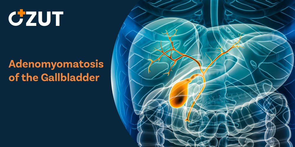
What Is Adenomyomatosis?
Adenomyomatosis occurs when the walls of the gallbladder become abnormally thickened. This thickening is accompanied by the formation of small pockets within the gallbladder tissue known as the company sinuses. These tiny outpouchings extend from the gallbladder lumen into the muscular wall layer.
The gallbladder is a small, pear-shaped organ located in the upper right portion of the abdomen. Its primary function is to store and concentrate bile produced by the liver. Bile plays an essential role in digestion by breaking down fats in the small intestine. When adenomyomatosis develops, the normal structure of the gallbladder wall becomes altered, though the organ typically continues to function.
Types of Adenomyomatosis
Medical professionals classify adenomyomatosis into four distinct types based on the location and extent of the gallbladder wall thickening:
- Local (Fundal) Adenomyomatosis : This is the most common type, involving thickening at a single site, typically at the fundus, which is the widest part of the gallbladder. Because of its location, it’s often referred to as fundal adenomyomatosis.
- Annular Adenomyomatosis : This type involves tissue thickening around the circumference of the gallbladder, creating an hourglass-like shape inside the organ.
- Segmental Adenomyomatosis : In this form, a larger area of tissue thickens, typically extending beyond the fundus. The affected portion of the gallbladder contracts inward while the remaining tissue appears normal.
- Diffuse Adenomyomatosis : This type affects the entire gallbladder, with widespread thickening throughout the organ.
Symptoms of Adenomyomatosis
Many individuals with adenomyomatosis experience no symptoms whatsoever. In fact, the condition is frequently discovered incidentally during medical tests conducted for other health concerns. This asymptomatic nature explains why many cases go undetected until imaging is performed for unrelated reasons.
When symptoms do occur, they may include:
- Upper right abdominal pain
- Discomfort after eating fatty foods
- Nausea
- Vomiting
- Symptoms similar to those of gallstones
It’s important to note that these symptoms aren’t exclusive to adenomyomatosis and could indicate other gallbladder conditions. Therefore, proper medical evaluation is essential for accurate diagnosis.
What Causes Adenomyomatosis?
Cellular Hyperplasia
Adenomyomatosis develops when cells in the gallbladder lining grow more rapidly than normal—a process known as hyperplasia. This accelerated growth leads to the thickening of the gallbladder wall and the formation of the characteristic the company sinuses.
Risk Factors
While the exact cause remains unclear, several factors may contribute to the development of adenomyomatosis:
- Increased pressure within the gallbladder
- Chronic inflammation
- Hormonal factors
- Aging (more common in middle-aged and older adults)
Research suggests that adenomyomatosis may be more prevalent in women than men, though it can affect individuals of any gender. The condition also appears to become more common with advancing age, with most diagnoses occurring in people over 50 years old.
Diagnosing Adenomyomatosis
Accurate diagnosis of adenomyomatosis is crucial, particularly to distinguish it from gallbladder cancer, which can sometimes present similarly on imaging tests. Several diagnostic approaches may be used:
Ultrasound
Ultrasound is typically the first imaging test used when gallbladder issues are suspected. During an ultrasound examination, radiologists look for characteristic findings of adenomyomatosis, including:
- Thickening of the gallbladder wall
- Presence of the company sinuses
- “Comet tail artifacts,” which are reflections of ultrasound waves off cholesterol crystals in the affected areas
Additional Imaging Tests
When ultrasound results are inconclusive or more detailed imaging is needed, healthcare providers may recommend:
- Magnetic Resonance Imaging (MRI) : MRI can provide detailed images of the gallbladder and surrounding tissues, helping to identify the characteristic features of adenomyomatosis.
- Computed Tomography (CT) Scan : CT scans may be used to evaluate the gallbladder and rule out other conditions.
- Endoscopic Ultrasound : This specialized procedure combines endoscopy and ultrasound to obtain detailed images of the digestive tract, including the gallbladder.
Biopsy
In cases where imaging tests cannot definitively distinguish between adenomyomatosis and gallbladder cancer, a biopsy may be necessary. This involves taking a small tissue sample from the gallbladder for microscopic examination. Gallbladder cancer is often aggressive and has a poor prognosis, making accurate differentiation between these conditions essential.
Treatment Options for Adenomyomatosis
Monitoring
If adenomyomatosis is confirmed and you don’t have any symptoms, treatment may not be necessary. Instead, your healthcare provider might recommend periodic monitoring with ultrasound examinations to track any changes in the condition over time.
Cholecystectomy (Gallbladder Removal)
For individuals experiencing symptoms related to adenomyomatosis, surgical removal of the gallbladder—known as cholecystectomy—may be recommended. This procedure is also considered when:
- Symptoms are persistent or severe
- There’s uncertainty about the diagnosis, and cancer cannot be completely ruled out
- The condition is accompanied by gallstones or other gallbladder issues
Cholecystectomy is typically performed laparoscopically, involving several small incisions rather than one large one. This minimally invasive approach generally results in shorter recovery times and less post-operative pain compared to traditional open surgery.
Medication
Currently, there are no medications specifically approved for treating adenomyomatosis. However, pain relievers may be prescribed to manage any discomfort associated with the condition.
Living Without a Gallbladder
The gallbladder is not an essential organ, and many people live normal, healthy lives after having it removed. Following cholecystectomy, bile flows directly from the liver into the small intestine rather than being stored in the gallbladder first.
After gallbladder removal, some individuals may need to make dietary adjustments, particularly in the early recovery period. These may include:
- Limiting fatty foods
- Eating smaller, more frequent meals
- Gradually reintroducing fiber into the diet
- Staying well-hydrated
Most people adapt to these changes within a few weeks or months after surgery, though some may experience longer-term digestive changes that require ongoing dietary management.
Prognosis and Outlook
The outlook for individuals with adenomyomatosis is generally favorable. Since the condition is benign, it doesn’t pose the same risks as cancerous conditions. Many people with adenomyomatosis never develop symptoms and require no treatment throughout their lives.
For those who undergo gallbladder removal due to symptomatic adenomyomatosis, the prognosis is typically excellent. Most individuals recover fully from surgery and can return to their normal activities within a few weeks.
Adenomyomatosis and Cancer Risk
While adenomyomatosis itself is not cancerous, some research has investigated whether there might be an association between long-standing adenomyomatosis and an increased risk of gallbladder cancer. However, current evidence does not strongly support a causal relationship between the two conditions.
Nevertheless, because adenomyomatosis can sometimes mimic the appearance of gallbladder cancer on imaging tests, thorough evaluation is essential to ensure an accurate diagnosis.
When to Seek Medical Attention
If you experience persistent pain in the upper right portion of your abdomen, particularly after meals, it’s advisable to consult a healthcare provider. While these symptoms could be related to various conditions, including adenomyomatosis, proper medical evaluation is necessary for accurate diagnosis and appropriate treatment.
Seek immediate medical attention if you experience:
- Severe abdominal pain
- Fever and chills
- Yellowing of the skin or eyes (jaundice)
- Dark urine or light-colored stools
These symptoms could indicate complications related to gallbladder disease or other serious conditions requiring prompt medical intervention.
Conclusion
Adenomyomatosis is a benign condition characterized by thickening of the gallbladder wall and the formation of the company sinuses. While often asymptomatic and discovered incidentally, it can sometimes cause discomfort and may require treatment, particularly when symptoms are present.
The condition’s ability to mimic gallbladder cancer on imaging tests makes accurate diagnosis crucial. With proper evaluation and appropriate management, individuals with adenomyomatosis can expect a favorable outcome, whether through monitoring of an asymptomatic condition or surgical intervention for symptomatic cases.
If you suspect you may have gallbladder issues, consulting with a healthcare provider is the first step toward diagnosis and developing an appropriate management plan tailored to your specific situation.