Lumbosacral Spine X-Ray: Purpose, Procedure, and What to Expect
A lumbosacral spine X-ray is an important diagnostic imaging test that allows healthcare providers to examine the bones of your lower back. Using minimal amounts of electromagnetic radiation, this procedure creates clear images that help medical professionals evaluate bone fractures, joint problems, and various other conditions affecting the lower spine region.
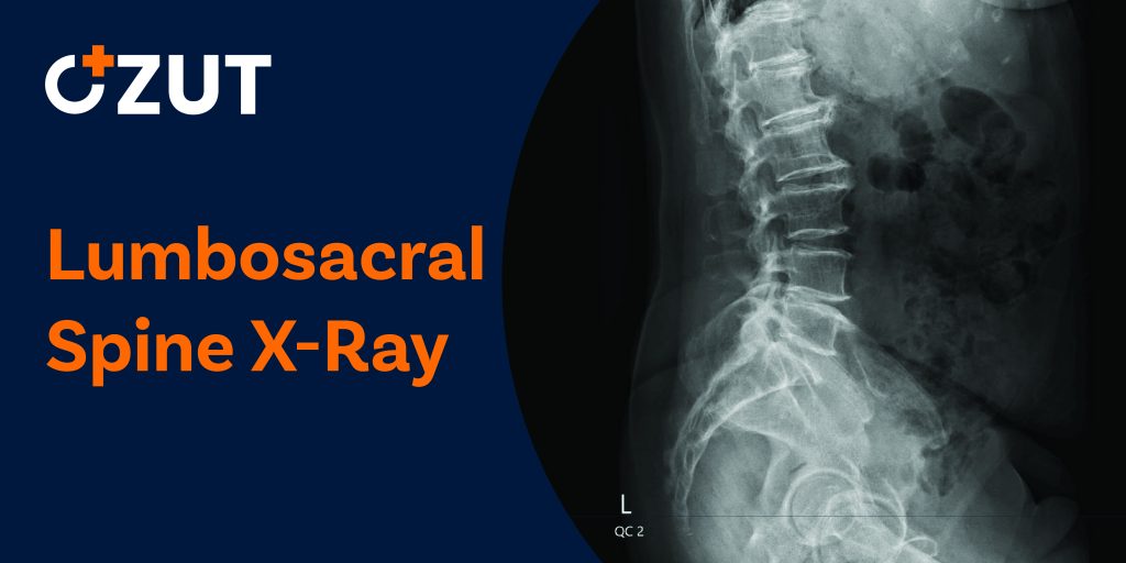
Understanding the Lumbosacral Spine
The spine is divided into several distinct sections, each serving specific functions in supporting the body and protecting the spinal cord. The lumbosacral spine specifically refers to the lower portions of the vertebral column.
Anatomy of the Lower Spine
The lumbosacral region consists of two primary components:
- Lumbar Spine : This is the lowest section of the spine, positioned below the thoracic (middle) and cervical (upper) spine segments. The lumbar region typically contains five vertebrae, labeled L1 through L5, and bears most of the body’s weight.
- Sacrum : This triangular-shaped bone acts as a protective shield at the back of your pelvis, situated directly below the lumbar spine. The sacrum connects the spine to the pelvis and helps transfer weight from the upper body to the lower limbs.
- Coccyx : Often called the tailbone, this small structure is located at the very bottom of the spine, just below the sacrum.
Understanding this anatomy helps explain why issues in the lumbosacral region often cause significant discomfort and mobility problems, as this area supports substantial weight and facilitates numerous movements.
Medical Reasons for Ordering a Lumbosacral X-Ray
Healthcare providers may recommend lumbosacral spine X-rays for various reasons. These imaging tests serve as valuable diagnostic tools for both acute injuries and chronic conditions affecting the lower back.
Diagnostic Applications
A lumbosacral X-ray can help identify or evaluate:
- Injuries from falls, accidents, or trauma
- Persistent or severe lower back pain
- Congenital conditions affecting spine development
- Osteoarthritis progression in spinal joints
- Advanced osteoporosis and related bone weakening
- Bone spurs (osteophytes) forming along vertebral edges
- Scoliosis or other abnormal spine curvatures
- Degenerative disc disease causing height loss in spinal discs
- Vertebral compression fractures
- Spinal alignment issues
Beyond initial diagnosis, these X-rays also help monitor disease progression and treatment effectiveness. For instance, a healthcare provider might order follow-up X-rays to assess how well a specific therapy is addressing osteoporosis or to track changes in spinal curvature over time.
Preparing for Your Lumbosacral X-Ray
Lumbosacral X-rays are routine, non-invasive procedures that require minimal preparation. However, following a few simple guidelines ensures the best possible imaging results and a smooth experience.
Before Your Appointment
Generally, no special preparation is needed before a lumbosacral X-ray. You can eat, drink, and take medications as usual unless specifically instructed otherwise by your healthcare provider. However, it’s always advisable to:
- Inform your doctor about any recent illnesses or medical conditions
- Mention any possibility of pregnancy, as special precautions may be necessary
- Provide a list of all medications, supplements, and vitamins you’re taking
- Discuss any previous reactions to contrast materials used in imaging procedures
What to Wear and Bring
On the day of your X-ray, consider wearing comfortable clothing without metal components. You’ll likely need to change into a hospital gown for the procedure, but comfortable attire makes the process easier. Remember to:
- Leave jewelry and accessories at home
- Wear clothes without zippers, buttons, or other metal fasteners if possible
- Bring your insurance information and identification
- Have a list of your current medications available
- Bring previous imaging studies if requested by your provider
Most importantly, inform the technologist about any metal implants from prior surgeries, such as pacemakers, artificial joints, or spinal hardware, as these can affect image quality and interpretation.
The Lumbosacral X-Ray Procedure
Understanding what happens during a lumbosacral X-ray can help ease any anxiety about the procedure. The process is straightforward and typically takes only a short time to complete.
Location and Equipment
Lumbosacral X-rays are performed in a hospital’s radiology department or at specialized diagnostic imaging centers. The main equipment includes:
- An X-ray machine with a large camera connected to an adjustable arm
- A flat table for the patient to lie on during certain views
- Digital or film receptors that capture the X-ray images
- Lead shields to protect parts of the body not being imaged
Positioning and Image Capture
The technologist will position you according to the specific views ordered by your healthcare provider. Many lumbosacral X-rays are performed while standing to evaluate how your body weight affects your spine alignment. This weight-bearing assessment provides valuable information about spinal stability and function under normal conditions.
Depending on what your doctor needs to see, you may be asked to assume several different positions during the examination:
- Standing position : You’ll stand with your back against the imaging plate for anteroposterior (front-to-back) and lateral (side) views
- Lying position : You may lie on your back, side, or stomach on the X-ray table
- Flexion and extension views : In some cases, images may be taken while you bend forward or arch backward to assess spinal mobility
- Oblique views : These angled images help visualize specific structures that might be obscured in standard views
For each image, the technologist will position the X-ray machine precisely, ask you to hold still, and sometimes instruct you to hold your breath for a few seconds while the image is captured. Remaining motionless during this brief moment is crucial for obtaining clear, diagnostic-quality images.
What to Expect During and After the Procedure
During the X-Ray
The actual X-ray exposure lasts only a fraction of a second for each image. You won’t feel anything when the X-ray is taken—there’s no pain, heat, or other sensation. The most challenging aspect for some patients is maintaining potentially uncomfortable positions or holding still if they’re experiencing back pain.
The entire procedure typically takes about 15 minutes, though this can vary depending on how many images are needed and whether any need to be retaken for clarity. The technologist will guide you through each position change and provide clear instructions throughout the process.
After Your X-Ray
Once all necessary images have been obtained, you can change back into your regular clothes if you were provided with a hospital gown. No recovery period is needed after a lumbosacral X-ray, and you can immediately resume your normal activities without restrictions.
The images will be reviewed by a radiologist—a physician specialized in interpreting medical images. The radiologist will analyze the X-rays and prepare a detailed report of their findings for your referring healthcare provider.
Getting Your Results
The timeframe for receiving results varies by facility. In some cases, preliminary results may be available the same day, while complete interpretations typically take 1-2 business days. Your healthcare provider will discuss the findings with you and explain what they mean for your diagnosis and treatment plan.
Based on the X-ray results, your doctor may:
- Make a definitive diagnosis
- Recommend additional imaging tests for more detailed views
- Suggest blood tests or other diagnostic procedures
- Develop a treatment plan for any identified conditions
- Schedule follow-up appointments to monitor your progress
Safety Considerations and Radiation Exposure
While X-rays do involve exposure to radiation, modern equipment and techniques have significantly reduced radiation doses compared to earlier technologies. For a standard lumbosacral spine X-ray, the radiation exposure is considered minimal and generally safe for most patients.
Radiation Safety Measures
Medical facilities follow strict protocols to ensure radiation safety:
- Using the lowest effective radiation dose necessary for diagnostic images
- Shielding areas of the body not being examined with lead aprons
- Carefully calibrating and maintaining X-ray equipment
- Following established guidelines for appropriate use of imaging
- Employing trained technologists who specialize in proper imaging techniques
Special Considerations
Certain populations require additional precautions when considering X-ray procedures:
- Pregnant women : X-rays are generally avoided during pregnancy unless absolutely necessary. If you are or might be pregnant, inform your healthcare provider before the procedure.
- Children : Since children are more sensitive to radiation effects, pediatric imaging protocols use adjusted settings to minimize exposure while still obtaining necessary diagnostic information.
- Frequent imaging : Patients requiring multiple X-rays over time should discuss radiation exposure concerns with their healthcare provider, who may suggest alternative imaging methods when appropriate.
For most adults, the benefits of accurate diagnosis through lumbosacral X-rays far outweigh the minimal risks associated with radiation exposure from a single examination.
Limitations and Alternative Imaging Methods
While lumbosacral X-rays provide valuable information about bone structures, they have certain limitations. X-rays primarily show bones and joints but provide limited visualization of soft tissues like muscles, ligaments, nerves, and intervertebral discs.
When Additional Imaging May Be Needed
Depending on your symptoms and the X-ray findings, your healthcare provider might recommend complementary imaging studies:
- MRI (Magnetic Resonance Imaging) : Provides detailed images of soft tissues, including spinal discs, nerves, and the spinal cord itself
- CT (Computed Tomography) : Offers cross-sectional images that can show more detail of bone structures and some soft tissues
- Myelogram : A specialized procedure combining contrast material with X-rays or CT scans to evaluate the spinal canal and nerve roots
- Bone scan : Uses a radioactive tracer to identify areas of increased bone activity that might indicate infection, fractures, or tumors
- Ult